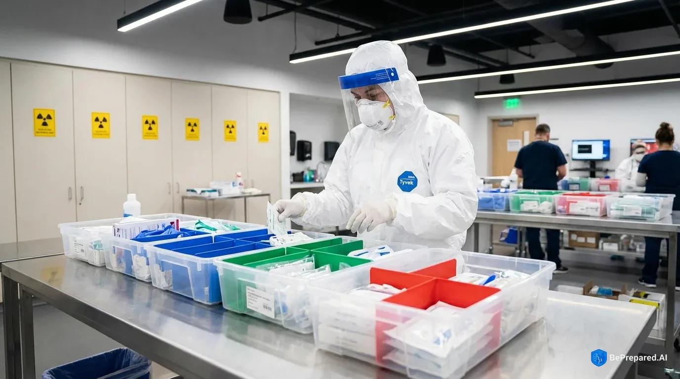
Understanding the Medical Challenges of Nuclear Events
When a nuclear detonation occurs, the medical response demands capabilities that extend far beyond conventional disaster preparedness. Whether the source is a weapon or a reactor accident, casualties arrive with a convergence of trauma types that simultaneously strain every aspect of medical care. Victims don't present with simple radiation exposure—they suffer blast injuries, thermal burns, traumatic wounds, and varying degrees of radiation contamination, all requiring immediate but contradictory treatment priorities.
The complexity begins with understanding that radiation injuries operate on a completely different timeline than conventional trauma. A person standing near a nuclear event might suffer severe burns and lacerations that demand immediate attention, yet the radiation exposure they received simultaneously won't manifest critical symptoms for hours or even days. This delayed presentation creates a triage nightmare. First responders and medical personnel must treat visible injuries while simultaneously assessing and documenting radiation exposure levels that will determine survival weeks later. The patient bleeding in front of you needs wound care now, but without proper radiation monitoring and decontamination protocols, you risk contaminating your medical supplies, your treatment area, and yourself—potentially rendering you unable to help anyone else.
Different radiation exposure scenarios create distinct medical challenges that require specific responses. External irradiation occurs when someone is exposed to radiation but doesn't carry radioactive particles on or in their body. Think of this like getting an X-ray—harmful while it's happening, but the person doesn't become radioactive afterward. These individuals need medical treatment for radiation sickness based on their exposure dose, but they don't require decontamination. Conversely, external contamination happens when radioactive particles settle on skin, clothing, or hair. These patients are essentially covered in a hazardous substance that continues emitting radiation, affecting both the victim and anyone nearby. They require thorough decontamination before entering medical facilities. The most severe scenario involves internal contamination, where radioactive materials enter the body through inhalation, ingestion, or open wounds. These particles irradiate tissues from the inside, requiring specialized pharmaceutical interventions like chelating agents to bind and remove the radioactive material from the body .
The timeline of medical needs following a nuclear event stretches across distinct phases, each presenting unique challenges. The immediate phase, spanning the first 24-48 hours, sees the highest casualty influx. Blast injuries, severe burns, and traumatic wounds dominate this period. Radiation exposure symptoms may not yet be apparent, but this window represents the critical opportunity for administering radiation countermeasures like potassium iodide and beginning decontamination procedures. The intermediate phase, from 48 hours to several weeks, brings the onset of acute radiation syndrome (ARS) in exposed individuals. Symptoms escalate from nausea and fatigue to bone marrow suppression, hemorrhaging, and immune system collapse. Patients who seemed stable during initial treatment may suddenly deteriorate as their radiation injuries manifest. The long-term phase extends months to years, dealing with cancer risks, organ damage, and psychological trauma .
Civilian medical infrastructure faces severe limitations in nuclear event response that preppers must understand and plan around. Most hospitals maintain virtually no radiation-specific medical supplies beyond basic detection equipment. The Strategic National Stockpile contains some radiation countermeasures, but distribution to affected areas could take 24-96 hours—far too late for time-sensitive interventions like potassium iodide administration, which must occur before or immediately after exposure to be effective . Medical facilities themselves become casualties in nuclear events. Hospitals within the blast radius sustain structural damage, while those outside face overwhelming patient surges that exceed capacity within hours. Staff shortages compound rapidly as healthcare workers either become casualties themselves or cannot reach facilities due to damaged infrastructure and contamination zones.
The electromagnetic pulse (EMP) from a nuclear detonation creates an often-overlooked medical challenge. Electronic medical equipment, digital medication dispensers, electronic health records, and communication systems can all fail simultaneously. Modern hospitals rely extensively on computerized systems for everything from medication administration to ventilator operation. An EMP transforms high-tech medical centers into pre-digital facilities, except staff haven't trained for manual operation of critical systems. Your personal medical preparedness must account for this reality—any radiation-specific medical equipment you stockpile must either function without electronics or include EMP-hardened alternatives.
The psychological dimension of radiation injuries complicates medical response in ways unique to nuclear events. Radiation carries a profound psychological terror because it's invisible, poorly understood by most people, and associated with slow, painful death. Medical personnel will face thousands of "worried well" individuals who fear exposure but have no actual injuries, overwhelming triage systems. Simultaneously, confirmed radiation casualties may refuse treatment out of hopelessness or fear of becoming pariahs. Healthcare providers themselves may refuse to treat contaminated patients, either from fear of exposure or family pressure to abandon their posts—a documented phenomenon in previous radiological incidents.
- Potassium Iodide (KI) Tablets
- 130mg tablets, 100-count bottle per person. Blocks radioactive iodine absorption. FDA-approved only.
- Decontamination Kit
- RSDL lotion, RadiacWash powder, disposable Tyvek suits, 5-gallon bucket system with filter
- Radiation Shielding Materials
- Lead sheets (2mm thickness), borated polyethylene panels, minimum 100 sq ft coverage area
Essential Radiation-Specific Medical Supplies
Building a comprehensive radiation medical supply cache requires understanding that conventional first aid supplies, while necessary, prove woefully inadequate for nuclear event scenarios. The specialized equipment needed to detect, monitor, and treat radiation exposure represents an entirely separate category of preparedness that most people overlook until it's too late.
Radiation detection and monitoring equipment forms the foundation of any radiation medical response. Without the ability to measure radiation levels, you're operating blind—unable to determine safe areas, assess exposure levels, or make informed treatment decisions. Personal radiation dosimeters should be your first acquisition. These devices measure accumulated radiation exposure over time, providing critical data about whether someone has received a dangerous dose. Modern electronic personal dosimeters (EPDs) offer real-time readings and alarm functions when exposure reaches preset thresholds, but they require batteries and are vulnerable to EMP effects. Backup passive dosimeters, such as thermoluminescent dosimeters (TLDs) or optically stimulated luminescence dosimeters (OSLs), don't provide immediate readings but survive EMP and require no power. You need both types—electronic for immediate decision-making and passive for reliable long-term monitoring .
Survey meters, often called Geiger counters, detect and measure radiation in the environment rather than accumulated personal exposure. These instruments identify contaminated areas, check whether decontamination was successful, and screen incoming casualties for external contamination. Professional-grade survey meters capable of detecting alpha, beta, and gamma radiation typically cost several thousand dollars, but they provide capabilities that cheaper consumer models lack. Alpha radiation, consisting of heavy particles that don't penetrate skin but cause severe damage if inhaled or ingested, requires specialized detectors that budget Geiger counters often can't detect. Given that internal contamination from alpha-emitting particles represents one of the most dangerous radiation scenarios, this detection capability isn't optional for serious preparedness .
Spectrometers take detection further by identifying specific radioactive isotopes present in contamination. This identification determines which pharmaceutical countermeasures to administer. Cesium-137 contamination requires different treatment than plutonium or cobalt-60 exposure. While spectrometers represent a significant investment—field-portable units can be quite expensive—they transform radiation response from educated guessing to targeted treatment. For group preparedness or community response teams, one quality spectrometer shared among members provides more value than multiple basic Geiger counters.
Decontamination supplies must be stockpiled in quantities that seem excessive until you understand the process. Effective decontamination removes 95-99% of external contamination, dramatically reducing ongoing radiation exposure and preventing internal contamination through wounds or ingestion . Water forms the basis of decontamination—substantial quantities of water. A single contaminated person may require significant water volumes for thorough decontamination, including multiple rinses. Your water storage calculations must account for this in addition to drinking, cooking, and sanitation needs. Decontamination doesn't require sterile water, but it should be clean enough to avoid introducing infections into wounds or abraded skin.
Specialized decontamination solutions prove more effective than water alone for certain contamination types. Calcium or zinc DTPA (diethylenetriaminepentaacetic acid) solutions can be used externally to bind radioactive particles before they absorb through skin. Household items like mild dish soap effectively remove many contaminants without damaging skin, but avoid harsh detergents or scrubbing that abrades skin—this drives contamination deeper into tissue rather than removing it. Medical-grade decontamination kits contain pH-neutral solutions specifically formulated to remove radioactive particles without causing tissue damage. Stock multiple kits, because you'll need separate supplies for each contaminated individual to prevent cross-contamination.
Decontamination infrastructure supplies often get overlooked. You need collection systems for contaminated runoff water, which itself becomes radioactive waste requiring containment. Heavy-duty plastic sheeting creates decontamination zones and channels runoff into collection barrels. Soft-bristle brushes, sponges (disposed after single use), and spray bottles allow controlled application of decontamination solutions. Scissors or trauma shears cut away contaminated clothing, which must be sealed in plastic bags marked as radioactive waste. The decontamination process generates substantial contaminated waste—plan for multiple 55-gallon drums with sealed lids for waste storage.
Radiation-specific pharmaceuticals represent the most time-sensitive and protocol-dependent supplies in your cache. Potassium iodide (KI) tablets receive the most attention in preparedness circles, but understanding their limitations proves as important as stockpiling them. KI protects only the thyroid gland from radioactive iodine isotopes, and only if taken before or immediately after exposure . The thyroid concentrates iodine from the bloodstream, and during a nuclear event, it can't distinguish between safe iodine and radioactive iodine-131. Taking KI floods the thyroid with stable iodine, preventing uptake of the radioactive version. This protection is valuable—thyroid cancer represents one of the most common radiation-induced malignancies—but KI does absolutely nothing for other radiation exposure effects.
Dosing protocols for KI vary by age because thyroid sensitivity to radiation decreases with age while KI side effects increase. Adults over 40 should only take KI if exposure exceeds 500 cGy to the thyroid—a threshold unlikely except very near a nuclear event. The recommended adult dose of KI is 130mg as a single dose when exposure is anticipated . Children require smaller doses: 65mg for ages 3-18, 32.5mg for ages 1 month to 3 years, and 16.25mg for newborns. These aren't daily doses—KI provides protection for approximately 24 hours per dose. Repeated daily dosing may be necessary if exposure continues, but extended KI use carries risks including thyroid suppression and allergic reactions . Stock KI in multiple dose sizes to avoid splitting tablets, which leads to dosing errors.
Prussian blue (ferric hexacyanoferrate) treats internal contamination from radioactive cesium and thallium by binding these elements in the intestines and preventing absorption. The bound radioactive material then passes through the digestive system rather than circulating through the body. Prussian blue reduces the biological half-life of cesium-137 from 110 days to about 30 days—a significant reduction in total radiation dose. The medication comes in 500mg capsules, with typical dosing at 3 grams three times daily for adults. Treatment continues until contamination levels drop to safe ranges, potentially requiring weeks of therapy. Prussian blue requires a prescription in the United States, making it difficult to stockpile, but its effectiveness for cesium contamination—one of the most common radioisotopes in nuclear events—makes it worth pursuing through preparedness-friendly physicians .
Calcium/zinc DTPA are essential for treating internal contamination with transuranics . These chelating agents bind to radioactive heavy metals (plutonium, americium, curium) and rare earth elements, forming compounds that the kidneys can filter and excrete. Without chelation therapy, these internally deposited radioactive materials continue irradiating tissues for years, virtually guaranteeing cancer and organ failure. DTPA must be administered as early as possible—effectiveness decreases significantly after 24 hours as radioactive particles incorporate into tissues. Initial treatment uses calcium-DTPA (1 gram via IV or nebulized inhalation), followed by zinc-DTPA for subsequent doses to avoid depleting the body's zinc and other essential minerals. Treatment may continue for months depending on contamination levels .
The challenge with DTPA is availability. It's manufactured in limited quantities, held primarily by government stockpiles, requires prescription and medical supervision, and must be administered IV or via nebulizer—not simple pill form. For serious preppers, particularly those with medical training, pursuing DTPA through official channels (occupational exposure programs, research institutions, or emergency preparedness grants) represents the only legal acquisition path. The alternative—accepting that internal contamination with transuranics likely means death—motivates some to extraordinary efforts to obtain this medication.
Filgrastim and other granulocyte colony-stimulating factors (G-CSF) treat the bone marrow suppression that occurs with acute radiation syndrome. Radiation damages rapidly dividing cells, and bone marrow cells divide constantly to produce blood cells. When radiation destroys bone marrow, white blood cell counts plummet, leaving victims vulnerable to overwhelming infections. G-CSF stimulates surviving bone marrow to increase white blood cell production, potentially preventing fatal infections during the critical weeks when the immune system is compromised. These medications require refrigeration, have limited shelf life (typically 2-3 years), and represent a significant financial investment. Multiple doses over 2-3 weeks may be needed, making this an expensive stockpile item, but for serious radiation exposure, it's potentially life-saving .
Advanced Trauma and Emergency Care Supplies
Nuclear events create a brutal convergence of injury types that demand comprehensive trauma supplies extending far beyond basic first aid kits. The blast wave, thermal pulse, and radiation exposure combine to produce casualties with multiple simultaneous injuries requiring advanced interventions. Get inside the nearest building immediately after a nuclear explosion to minimize exposure to the initial radiation pulse and subsequent fallout.
Burn Treatment Supplies for Radiation-Complicated Injuries:
Thermal burns from the nuclear flash affect exposed skin surfaces, often covering extensive body areas. Radiation exposure complicates burn healing by suppressing immune function and impairing tissue regeneration. Your burn supply cache must address both immediate stabilization and extended wound management.
Sterile burn dressings in multiple sizes (4x4, 8x10, 12x18 inch) with non-adherent surfaces prevent the dressing from bonding to damaged tissue. Removing adherent dressings tears away healing tissue and causes excruciating pain. Silver sulfadiazine cream (Silvadene) serves as the standard topical antimicrobial for burn care. Burns create ideal bacterial growth conditions—warm, moist, nutrient-rich environments. Silvadene prevents infection while promoting healing. Stock this in bulk quantities because application requires multiple daily treatments over weeks.
Hydrogel burn dressings cool tissue and maintain moisture balance while preventing infection. These dressings absorb wound exudate while keeping the burn surface hydrated—critical for healing. Elastic conforming gauze secures burn dressings without constricting circulation as swelling develops. Burns cause significant tissue swelling in the hours following injury. Circumferential dressings that fit properly initially can become tourniquets as swelling progresses.
Burn sheets (large sterile sheets) cover extensive body surface area burns during transport or initial treatment. A person with 40% body surface area burns can't be covered with standard 4x4 dressings—you need large-format sterile coverings. Fluid replacement solutions including lactated Ringer's and normal saline in IV bags address the massive fluid losses through damaged skin. Burn victims can lose liters of fluid daily through their wounds, leading to hypovolemic shock without aggressive replacement.
Specialized burn calculation tools (Lund-Browder charts) assess total body surface area affected and calculate fluid replacement needs. The "rule of nines" provides rough estimates, but Lund-Browder charts account for age-related body proportion differences and provide more accurate assessments. Pain management medications including injectable morphine or fentanyl control burn pain that exceeds what oral medications can manage. Severe burns produce pain that breaks through oral analgesics. Injectable opioids provide the only adequate relief.
Antibiotic ointments beyond standard triple-antibiotic, including bacitracin and mupirocin, combat resistant organisms that colonize burn wounds. Pseudomonas and MRSA commonly infect burns, requiring targeted antimicrobials. Sterile irrigation solutions in large volumes (liter bags) clean burns before dressing application. Proper burn care requires copious irrigation to remove debris and dead tissue before applying antimicrobials and dressings.
Blast Injury Trauma Supplies:
The blast wave from a nuclear detonation creates overpressure that tears tissue, ruptures organs, and drives fragments into the body. Blast injuries combine blunt trauma, penetrating wounds, and internal organ damage in patterns rarely seen outside military combat.
Hemostatic gauze (Combat Gauze, Celox, QuikClot) controls severe bleeding from blast fragmentation wounds. These gauze products contain agents that accelerate clotting, stopping hemorrhage that pressure alone can't control. Stock at least 10-20 packages because single wounds may require multiple applications. A deep fragmentation wound might need three or four packages of hemostatic gauze packed into the wound cavity to achieve hemostasis.
Tourniquets (CAT or SOFTT-W models) in quantities of 4-6 address complex extremity injuries. Blast victims may require multiple tourniquets—one per injured limb. Modern windlass tourniquets can be self-applied and maintain pressure for hours without tissue damage when properly used. Chest seals (both vented and non-vented types) treat pneumothorax from blast overpressure. Blast lungs represent a common life-threatening injury. The pressure wave ruptures alveoli, causing air leaks into the pleural space that collapse the lung. Chest seals prevent air entry through chest wounds while allowing trapped air to escape.
Nasopharyngeal airways in multiple sizes maintain airways in unconscious blast victims. Blast injuries often cause altered consciousness from traumatic brain injury. Airway adjuncts prevent tongue obstruction and maintain ventilation. Needle decompression kits (14-gauge, 3.25-inch angiocatheters) provide emergency tension pneumothorax treatment. When air trapped in the pleural space creates pressure that shifts the mediastinum and impairs cardiac function, needle decompression releases the pressure and saves the patient's life.
Emergency cricothyrotomy kits establish airways when facial trauma prevents other methods. Blast injuries to the face can make oral or nasal intubation impossible. Cricothyrotomy creates a surgical airway through the cricothyroid membrane, bypassing upper airway obstruction. Israeli bandages or emergency trauma dressings with built-in pressure applicators control extremity hemorrhage. These dressings combine absorbent pads with pressure bars and elastic wraps in a single package, allowing rapid application even by the injured person.
Occlusive dressings in various sizes seal penetrating chest or abdominal wounds. Any wound that penetrates the thoracic or abdominal cavity requires occlusive coverage to prevent air entry and organ herniation. Splinting materials including SAM splints, air splints, and traction splints address complex fractures from structural collapse. Blast victims trapped under rubble sustain crush injuries and fractures requiring immobilization before and during transport.
Cervical collars in multiple sizes protect spinal injuries. Blast victims often sustain spinal injuries from being thrown by overpressure or struck by debris. Spinal precautions prevent converting incomplete cord injuries into complete paralysis through improper handling.
Advanced Wound Care for Contaminated Environments:
Radiation contamination transforms routine wound care into complex decontamination procedures. Wounds provide direct pathways for radioactive particles to enter the bloodstream, making contaminated wounds medical emergencies requiring immediate specialized treatment.
Surgical irrigation systems with splash shields enable high-volume wound cleaning. Contaminated wounds require aggressive irrigation with liters of solution to flush out radioactive particles before they absorb into tissue. Standard squeeze bottles lack the pressure and volume capacity for adequate irrigation. Sterile surgical instruments including forceps, scissors, scalpels, and hemostats allow wound exploration and debridement. Contaminated wounds may contain embedded radioactive fragments requiring surgical removal.
Suture kits with multiple suture types (absorbable and non-absorbable, various gauges) close wounds after decontamination confirms no residual radioactive particles. You can't close a contaminated wound—doing so traps radioactive material inside where it will irradiate tissue continuously. Wound vacuum systems (negative pressure wound therapy) manage large wounds that can't be immediately closed. These systems apply controlled suction to wounds, removing fluid, reducing edema, and promoting granulation tissue formation.
Antimicrobial dressings containing silver or iodine combat infected wounds in radiation-immunocompromised patients. Radiation exposure suppresses immune function, making infection nearly inevitable in contaminated wounds. Antimicrobial dressings provide continuous local antimicrobial activity. Sterile gloves in massive quantities (boxes of 100+) prevent cross-contamination. Every patient contact requires fresh gloves. Contaminated gloves transfer radioactive particles between patients, contaminating clean wounds and spreading contamination throughout your treatment area.
Sterile drapes create clean fields during procedures in less-than-ideal environments. When you're performing wound care in a garage or basement, sterile drapes establish a controlled sterile zone around the surgical site. Betadine or chlorhexidine in gallon containers provide repeated wound cleaning and skin preparation. You'll use enormous quantities of surgical prep solution when treating multiple contaminated casualties.
Sterile saline in large volumes (case quantities of liter bottles) serves irrigation and wound cleaning needs. A single contaminated wound might require 2-3 liters of irrigation. Multiple casualties exhaust small stockpiles rapidly. Wound packing materials including sterile gauze strips manage deep wound cavities. Deep wounds can't be closed immediately—they require packing to control bleeding and prevent abscess formation while healing from the inside out.
Emergency Surgical Supplies:
Severe blast and burn injuries may require emergency surgical interventions that can't wait for hospital care. When hospitals are destroyed, overwhelmed, or unreachable, your medical cache must include basic surgical capabilities.
Scalpel handles and disposable blades in multiple sizes (#10, #11, #15 blades) perform emergency procedures. Different blade shapes serve different purposes—#10 for large incisions, #11 for precise cuts, #15 for delicate work. Hemostats and clamps in various sizes control bleeding during emergency surgery. You need multiple hemostats because complex wounds may require a dozen or more clamps simultaneously controlling different bleeding vessels.
Retractors hold tissue aside during wound exploration or emergency procedures. You can't perform surgery on deep wounds without retractors to maintain visualization of the surgical field. Surgical drains (Penrose and Jackson-Pratt types) manage fluid accumulation in wounds. Large wounds and surgical sites accumulate blood and serum that must drain to prevent infection and promote healing.
Chest tube insertion kits with tubes in multiple sizes treat pneumothorax and hemothorax. Blast injuries commonly cause air or blood accumulation in the pleural space. Chest tubes drain these collections and allow lung re-expansion. Sterile surgical gowns and drapes maintain aseptic technique during procedures. Surgery in field conditions already carries high infection risk—proper sterile technique is your only defense against overwhelming wound infections.
Surgical staple guns enable rapid wound closure in mass casualty situations. Staples close wounds faster than sutures, though with less precision. When you have ten casualties waiting and limited time, staples provide adequate closure with acceptable cosmetic results. Bone saws and orthopedic equipment manage crush injuries and potential amputations. Severe crush injuries with vascular compromise may require amputation to save the patient's life. Without proper orthopedic instruments, you can't perform these procedures.
Catheterization supplies including Foley catheters in multiple sizes manage urinary retention in immobile patients. Blast and burn victims often can't urinate normally due to injury, shock, or immobility. Bladder distension causes severe pain and can lead to rupture. IV start kits with multiple catheter sizes, plus central line kits, establish vascular access for patients requiring extended IV therapy. Burn victims and radiation casualties need IV access for days or weeks. Peripheral IVs fail after 72-96 hours, requiring central lines for continued access.
Medication Stockpile for Trauma Management:
Pharmaceutical supplies transform your trauma equipment from wound dressing capability into actual medical treatment. Without medications, you can bandage injuries but can't treat infection, control pain, or manage the systemic effects of trauma and radiation.
Broad-spectrum antibiotics (cephalosporins, fluoroquinolones, carbapenems) in both oral and IV formulations prevent and treat infections. Radiation-damaged immune systems make infection nearly certain in any wound. Early aggressive antibiotic therapy prevents minor infections from becoming fatal sepsis. Stock multiple antibiotic classes because resistant organisms require alternative agents.
Pain management medications ranging from acetaminophen through tramadol to morphine and fentanyl provide escalating pain control. Different injuries require different analgesic potency. Minor wounds respond to acetaminophen, moderate injuries need tramadol or hydrocodone, and severe burns or blast injuries require morphine or fentanyl. Stock the full spectrum to match medication to injury severity.
Antiemetics (ondansetron, promethazine) in high quantities manage radiation-induced nausea. Radiation sickness causes severe, persistent nausea and vomiting that leads to dehydration and prevents oral medication and nutrition. Antiemetics don't cure radiation sickness, but they make patients comfortable enough to maintain hydration and nutrition.
Antidiarrheal medications (loperamide, bismuth subsalicylate) manage radiation-induced gastrointestinal damage. Radiation destroys the rapidly dividing cells lining the intestines, causing severe diarrhea. Fluid losses from radiation-induced diarrhea can cause fatal dehydration. Antidiarrheals slow intestinal motility and reduce fluid losses.
Corticosteroids (dexamethasone, prednisone) manage inflammation and certain radiation effects. Steroids reduce cerebral edema from traumatic brain injury, treat some radiation-induced inflammatory responses, and provide symptomatic relief for various trauma complications. Anticoagulants and clotting factors address the coagulation abnormalities radiation causes. Radiation can cause both bleeding disorders (through platelet destruction) and dangerous clotting (through vascular damage). You need medications to address both extremes.
Electrolyte replacement solutions correct the severe imbalances that occur with radiation sickness. Vomiting, diarrhea, and kidney damage from radiation cause sodium, potassium, and other electrolyte derangements that can cause cardiac arrhythmias and death. Anticonvulsants prevent and treat seizures from radiation exposure and traumatic brain injuries. Some radiation exposures and head trauma cause seizures requiring medication control.
Sedatives manage the psychological trauma and agitation common in nuclear event casualties. The terror of radiation exposure, pain from injuries, and loss of loved ones create severe psychological distress. Sedatives provide symptomatic relief and allow patients to rest and heal.
Blood and Fluid Management:
Trauma and radiation injuries cause massive fluid shifts requiring aggressive replacement. Without adequate fluid management capabilities, patients die from hypovolemic shock despite successful wound treatment.
IV administration sets with multiple drip chambers and roller clamps, plus pressure bags for rapid infusion, deliver fluids at rates matching patient needs. Slow drip rates suffice for maintenance fluids, but hemorrhagic shock requires rapid infusion under pressure. Blood collection and transfusion supplies enable emergency transfusions if you have capability and training. Direct person-to-person transfusion can save lives when stored blood isn't available, though it carries significant risks.
Plasma volume expanders (albumin, hetastarch) treat shock when blood products aren't available. These solutions remain in the vascular space longer than crystalloid solutions, providing better hemodynamic support. Electrolyte solutions in variety (normal saline, lactated Ringer's, D5W) address different clinical situations. Normal saline for simple volume replacement, lactated Ringer's for burn resuscitation, D5W for hypoglycemia.
Blood pressure cuffs and stethoscopes monitor hemodynamic status. You can't manage shock without blood pressure monitoring. Automated BP cuffs require batteries and are EMP-vulnerable—manual cuffs and stethoscopes provide EMP-proof monitoring. Pulse oximeters (preferably with spare batteries or hand-crank charging) monitor oxygen saturation. Blast lung injuries and smoke inhalation cause hypoxemia requiring oxygen therapy. Pulse oximetry guides oxygen administration.
Blood glucose monitoring systems with extensive test strip supplies manage diabetic patients and stress hyperglycemia. Trauma and radiation exposure cause blood sugar elevations even in non-diabetics. Diabetic patients face life-threatening glucose swings during disasters. Glucose monitoring prevents both hypoglycemic coma and diabetic ketoacidosis.
PPE and Contamination Control
Protecting yourself while treating contaminated casualties requires understanding that radiation PPE serves a fundamentally different purpose than biological or chemical protection. You're not preventing radiation from penetrating your body—most PPE materials don't stop gamma radiation. Instead, you're preventing radioactive particles from contaminating your skin, being inhaled into your lungs, or being ingested. This distinction shapes every aspect of your PPE approach. Minimum PPE requirements include N95 masks, disposable gowns, and multiple pairs of gloves .
Step 1: Establish Your Baseline Protection Layer
Begin with a full-body coverall made from non-porous material like Tyvek or similar spun polyethylene. These suits prevent radioactive dust and particles from reaching your skin or clothing underneath. Select coveralls with attached hoods and elastic wrist and ankle closures to minimize gaps. The material should be thick enough to resist tears (Tyvek 1422A or heavier), as compromised suits provide false security. Before donning the coverall, dress in dedicated "contamination zone" scrubs or clothing that you'll dispose of or thoroughly decontaminate afterward. Never wear your regular clothing under contamination PPE—cross-contamination through tears or during removal makes your everyday clothes radioactive waste.
Over the coverall, add a disposable apron made from heavy-duty plastic or rubberized material. This seems redundant, but the apron takes the brunt of contamination during patient care, particularly during decontamination procedures where you're directly handling contaminated clothing and washing contaminated skin. When the apron becomes heavily contaminated, you can remove and replace it without doffing your entire suit, extending your working time in the contamination zone. Secure the apron ties outside your coverall so you can reach them with gloved hands during removal.
Step 2: Implement Respiratory Protection
Respiratory protection represents your most critical PPE component because inhaled radioactive particles cause internal contamination that's difficult or impossible to treat. A properly fitted N95 respirator provides minimum acceptable protection, filtering a high percentage of airborne particles. However, N95s offer no margin for error. Improper fit, beard growth preventing seal, or extended use degrading the filter all compromise protection. Fit-test your N95s before emergencies, and stock multiple sizes to accommodate different facial structures.
P100 respirators provide superior protection, filtering an even higher percentage of airborne particles. Full-face P100 respirators add eye protection and prevent face-touching, which commonly transfers contamination to mucous membranes. The disadvantage is communication difficulty and the need for training to prevent panic from the claustrophobic sensation. Practice wearing your respirator for extended periods—if you can't tolerate 30 minutes during training, you won't function effectively during an actual event when you might need to wear it for hours.
Powered Air-Purifying Respirators (PAPRs) represent the gold standard for radiation work. These systems use battery-powered fans to draw air through HEPA filters, providing positive pressure inside the hood that prevents contamination entry even if the seal isn't perfect. PAPRs are more comfortable for extended wear, don't require fit testing, and work with beards. The tradeoffs include significant financial investment, battery dependence, and the bulk of the system. For serious preparedness groups pooling resources, several PAPR units with spare batteries and filters provide capabilities that individual N95 stockpiles can't match.
Step 3: Layer Your Hand Protection
Double-gloving represents standard practice in radiation contamination scenarios. Start with a surgical glove as your inner layer, ensuring proper fit without excess material that creates fumbling. This inner glove stays on throughout your time in the contamination zone, providing continuous protection. The outer glove—a heavier nitrile or neoprene examination glove—gets changed frequently as it becomes contaminated during patient care. This layering allows you to remove grossly contaminated outer gloves without exposing your skin, then continue working with the clean inner glove until you can don fresh outer gloves.
Extend your gloves over the wrist cuffs of your coverall, then secure them with medical tape. This overlap prevents contamination from running down your gloves onto your wrists during decontamination procedures. Use brightly colored tape so you can visually confirm seal integrity. Some practitioners add a third layer of heavy-duty work gloves over the medical gloves when handling sharp debris or during physically demanding decontamination work, removing these work gloves before performing detailed medical procedures.
Step 4: Protect Your Feet and Lower Legs
Boot covers made from heavy-duty plastic or rubberized material go over your shoes, extending to mid-calf or knee height. Secure boot covers with tape around your calves, over the ankle cuffs of your coverall. Your feet will walk through contaminated water during decontamination, step in contaminated debris, and generally contact the most contaminated surfaces in your work area. Plan to change boot covers multiple times during extended operations, and have a clean path established where you can step out of contaminated covers into clean ones without tracking contamination.
Dedicated rubber boots worn instead of boot covers provide more durable protection for extended operations. These boots stay in your contamination zone and never enter clean areas. The disadvantage is the need to decontaminate them thoroughly rather than simply disposing of covers, but for multi-day operations, the durability justifies this tradeoff.
Step 5: Add Eye and Face Protection
Safety goggles with indirect venting prevent contaminated particles from reaching your eyes while allowing air circulation to prevent fogging. Direct-vent goggles or those with no venting at all create unbearable fogging during physical work. If you're using a half-face respirator rather than full-face, goggles become mandatory. Face shields worn over goggles add splash protection during decontamination procedures when you're spraying water and solutions that may contain dissolved radioactive particles. The shield protects your respirator from gross contamination, extending its usable life.
Step 6: Create Contamination Control Zones
Establish three distinct zones before beginning contamination response operations. The "hot zone" contains confirmed contamination—patients arrive here, decontamination occurs here, and everything in this zone is considered contaminated. The "warm zone" serves as a transition area where you remove contaminated PPE, perform personnel monitoring, and stage supplies moving into the hot zone. The "cold zone" is your clean area—no contaminated materials, no contaminated PPE, and where you rest, eat, and perform medical procedures on decontaminated patients.
Mark these zones clearly with colored tape, cones, or rope barriers. Traffic flows one direction: contaminated patients and materials move from hot to warm to cold zones. Clean supplies move from cold to warm to hot zones. People never move directly from hot to cold zones without passing through warm zone decontamination. This seems obvious during training, but during high-stress operations with multiple casualties, zone discipline breaks down rapidly without clear visual markers and designated zone monitors who do nothing but enforce traffic patterns.
Step 7: Implement PPE Removal Protocols
Doffing contaminated PPE without contaminating yourself requires deliberate, practiced technique. The fundamental principle: assume the outside of everything is contaminated, and never touch contaminated surfaces with bare skin or clean gloves. Begin removal in the warm zone with the most contaminated items—outer gloves and apron. Have a partner monitor your technique and use a survey meter to check for contamination transfer.
Remove outer gloves using the glove-in-glove technique: pinch the outside of one glove near the wrist and peel it off, turning it inside out. Hold this removed glove in your still-gloved hand. Insert fingers of your ungloved hand inside the wrist of the remaining glove, touching only the clean interior surface, and peel it off inside-out over the first glove. Both gloves are now inside-out, contained, and only clean surfaces contacted your skin. Immediately don fresh outer gloves before continuing—you still need hand protection for removing the rest of your PPE.
Remove your apron by breaking or cutting the ties (never pull contaminated ties over your head), rolling it inward on itself to contain contamination, and disposing it in radioactive waste containers. Remove boot covers by stepping out of them onto a clean surface—never sit down and handle them with your hands. Remove your coverall by peeling it forward and down, turning it inside out as you go, keeping contaminated surfaces contained. Finally, remove your respirator by handling only the straps, never touching the filter surface, and dispose it in waste containers unless you're using a PAPR that will be decontaminated.
After PPE removal, perform whole-body monitoring with a survey meter, paying particular attention to hands, feet, and head—the areas most likely to have contamination breakthrough. If monitoring reveals contamination, immediately perform personal decontamination before entering the cold zone. This monitoring step is non-negotiable—skipping it means potentially bringing contamination into your clean area, contaminating all your supplies, equipment, and personnel.
Step 8: Decontaminate Medical Equipment
Medical equipment used in contaminated areas requires systematic decontamination before returning to supply. Survey all equipment with radiation detectors before beginning decontamination to establish baseline contamination levels. Equipment with only surface contamination can be decontaminated through washing with soap and water, followed by monitoring to confirm contamination removal. Equipment with contamination in crevices or porous materials may require specialized cleaning or disposal as radioactive waste.
Establish a dedicated equipment decontamination station in your warm zone with wash basins, brushes, cleaning solutions, and rinse water. Clean items move from dirty side to clean side of the station, with monitoring between stages. Equipment that cannot be adequately decontaminated gets sealed in plastic bags, labeled with radiation symbols and contamination levels, and stored as radioactive waste. This seems wasteful, but using contaminated equipment in your clean treatment area defeats the entire purpose of zone control.
Storage and Maintenance Protocols
The most comprehensive radiation medical supply cache becomes worthless if materials degrade, pharmaceuticals expire, or equipment malfunctions during the crisis. Environmental control, systematic rotation, and rigorous documentation transform a static stockpile into a reliable medical capability.
Radiation pharmaceuticals demand the strictest storage conditions in your cache. Potassium iodide tablets can remain stable for several years when stored properly, but improper storage dramatically shortens this lifespan. KI requires cool (59-77°F), dry conditions away from light. Heat and humidity cause tablets to degrade, turning them yellow or brown—a visual indicator of reduced potency. Store KI in original packaging or transfer to amber glass bottles with desiccant packets, never loose in plastic bags where moisture accumulation destroys potency. Date all KI containers with acquisition date and expiration date, and inspect quarterly for discoloration. Even expired KI retains partial effectiveness, so don't discard expired tablets—segregate them as "second-tier" supplies for use if fresh stocks are exhausted .
Prussian blue capsules require similar cool, dry storage but have the additional complication of limited availability and prescription status. The 500mg capsules maintain potency for 2-3 years under proper conditions. Given the difficulty of acquiring Prussian blue, implement a strict first-in-first-out rotation system. As expiration approaches, some preppers consume expiring capsules prophylactically (Prussian blue has low toxicity and is used for other medical conditions) and replace them with fresh stock, though this requires physician consultation. Alternatively, establish relationships with other preparedness groups to trade expiring supplies—your Prussian blue expiring in six months might have years of life remaining for someone who acquired theirs earlier.
DTPA products present the most challenging storage requirements. Calcium-DTPA and zinc-DTPA are typically supplied as solutions in vials or ampules requiring refrigeration at 36-46°F. Freezing damages the solution, while temperatures above 46°F accelerate degradation. This refrigeration dependency creates vulnerability—power outages from EMP or infrastructure damage eliminate refrigeration exactly when you need these medications most. Address this through multiple strategies: maintain a backup power source (generator or solar system) dedicated to medical refrigeration, store DTPA in a high-quality cooler with frozen gel packs that maintain temperature for 48-72 hours during power outages, and if you have access to multiple DTPA sources, keep one refrigerated cache at your primary location and another at a distant backup location with independent power.
Filgrastim and other G-CSF medications similarly require refrigeration and have limited shelf life (24-36 months). These medications represent a substantial financial investment, making expiration-related waste financially painful. Rotate G-CSF supplies by establishing relationships with cancer treatment centers or veterinary clinics that use these medications regularly. As your stockpile approaches expiration, donate it to these facilities (they can often use medications within months of expiration) and replace it with fresh stock. This rotation provides a tax deduction for the donation while ensuring your cache remains viable.
Radiation detection equipment requires regular testing and calibration to ensure accuracy when needed. Electronic personal dosimeters and survey meters should be function-tested monthly—turn them on, verify battery levels, and confirm the device responds to a check source. Check sources are small, sealed radioactive samples (often cesium-137 or cobalt-60) that emit known radiation levels for calibrating and testing detection equipment. Store your check source in a shielded container (lead pig) away from your dosimeters to prevent false accumulated dose readings. Annual calibration by qualified technicians ensures your meters provide accurate readings. Uncalibrated meters may read significantly high or low, leading to dangerous decisions based on false data .
Passive dosimeters (TLDs and OSLs) require professional reading after exposure—you can't check them yourself. However, you can verify their physical integrity. Inspect dosimeters quarterly for cracks, moisture intrusion, or physical damage. Store passive dosimeters in their original packaging away from radiation sources, heat, and light. Maintain a log of each dosimeter's serial number and issue date. Some preppers send one dosimeter from their batch to a reading service annually as a control check—if it shows unexpected exposure, it indicates storage problems affecting the entire batch.
Decontamination supplies need less stringent environmental control but require quantity monitoring. Your decontamination water supply should be tested annually for contamination and algae growth. Rotate water supplies every 1-2 years, using old stocks for irrigation or other non-critical purposes while replacing with fresh water. Decontamination solutions (chelating agents, specialized soaps) have shelf lives of 2-5 years depending on formulation. Inspect solution containers quarterly for leaks, swelling, or discoloration indicating degradation. Plastic sheeting, collection barrels, and other decontamination infrastructure supplies have indefinite shelf life but should be inspected annually for rodent damage, UV degradation (if stored where sunlight reaches), or deterioration.
Trauma supplies and PPE require systematic rotation based on varying shelf lives. Sterile medical supplies (gauze, bandages, surgical instruments) remain sterile as long as packaging integrity is maintained. Inspect sterile packages quarterly for tears, moisture intrusion, or package swelling. Damaged packages lose sterility—repackage the contents in heat-seal bags with sterilization (autoclave or chemical sterilization if you have capability) or downgrade to "clean but non-sterile" supplies for use in less critical applications. Hemostatic agents like Combat Gauze have 3-5 year shelf lives—date these clearly and rotate approaching expiration to your everyday first aid kits while moving fresh stock into your radiation cache.
Medications in your trauma supply require the most aggressive rotation. Antibiotics, pain medications, and other pharmaceuticals typically have 2-5 year shelf lives, though many remain effective for years beyond expiration dates when stored properly. The military's Shelf Life Extension Program found that 90% of medications remain effective 5-10 years past expiration when stored in cool, dry conditions. However, certain medications—liquid antibiotics, epinephrine, insulin—degrade more rapidly and should be rotated at expiration. Implement a medication tracking spreadsheet documenting acquisition date, expiration date, quantity, and storage location for every pharmaceutical in your cache. Set quarterly reminders to review medications expiring in the next 6-12 months, allowing time to use or donate approaching-expiration supplies while acquiring replacements.
Documentation systems transform your supply cache from a pile of equipment into an organized medical capability. Create a master inventory spreadsheet listing every item, its quantity, storage location, acquisition date, expiration date (if applicable), and last inspection date. Update this inventory quarterly during your inspection cycle. Photograph your storage area and individual supply categories—these photos help you quickly locate items during high-stress events and provide documentation for insurance purposes if your cache is damaged or destroyed. Print copies of your inventory and store them in multiple locations (with your supplies, at a backup location, and in your evacuation kit)—digital-only records become inaccessible during power outages or EMP events.
Standard operating procedures (SOPs) for your radiation medical supplies should be written, practiced, and stored with your equipment. Create laminated quick-reference cards for critical procedures: KI dosing by age, DTPA administration protocols, decontamination procedures, PPE donning and doffing sequences. These cards provide guidance when stress and fatigue impair memory. Include source citations on your cards—during a crisis, you may need to justify your protocols to other responders or convince reluctant patients to accept treatment. "The CDC recommends this dose" carries more weight than "I think you should take this."
Training schedules ensure you can actually use your supplies effectively. Quarterly practice sessions should cover radiation detection equipment operation, PPE donning and doffing, decontamination procedures, and trauma care skills. Annual training exercises simulate complete scenarios—contaminated casualty arrival, decontamination, treatment, and waste management. Video record training sessions to identify mistakes and track improvement. Involve everyone who might participate in your radiation medical response—a cache maintained by one person becomes useless if that person is incapacitated during the event.
Environmental monitoring of your storage area provides early warning of conditions that might damage supplies. Install temperature and humidity sensors (data-logging models record highs and lows over time) in your storage area. Ideal conditions maintain 60-70°F and 30-50% relative humidity. Temperature spikes indicate HVAC failures requiring immediate attention. Humidity above 60% promotes mold growth and medication degradation—address through dehumidifiers or improved ventilation. Pest control is essential—rodents chew through packaging and contaminate supplies with droppings. Quarterly pest inspections and preventive measures (sealed containers, traps, exclusion methods) protect your investment.
Security measures protect your radiation medical supplies from theft while maintaining accessibility during emergencies. The challenge is that these supplies have high value—both monetary and survival value—making them theft targets if their existence becomes known. Store supplies in locked cabinets or rooms, but ensure multiple trusted individuals have access codes or keys. A cache that only one person can access becomes worthless if that person is killed or incapacitated during the event. Consider distributing your cache across multiple locations—primary cache at your main residence, backup cache at a secondary location, and grab-and-go kit in your vehicle. This distribution prevents total loss if one location is destroyed or inaccessible.
The financial investment in radiation medical supplies is substantial—a comprehensive cache can represent a significant expenditure depending on depth and group size. Implement a systematic acquisition plan rather than attempting to purchase everything immediately. Prioritize purchases based on likelihood of use and time-sensitivity: radiation detection equipment and KI first (immediately useful and time-critical), followed by decontamination supplies and PPE, then trauma supplies, and finally the hardest-to-obtain pharmaceuticals like DTPA and G-CSF. Spread purchases over months or years to distribute costs and allow time for learning about each category before moving to the next. Join group purchases with other preparedness-minded individuals to access bulk pricing and share expensive items like spectrometers that don't need individual ownership.
Insurance and legal considerations surround radiation medical supply stockpiling. Homeowner's insurance may not cover large medical supply caches—verify coverage limits and consider additional riders for high-value stockpiles. Prescription medications legally require prescriptions—work with preparedness-friendly physicians who understand the need for emergency supplies. Some medications (particularly opioid pain relievers and certain antibiotics) face legal restrictions on quantity prescribed. Document everything: prescriptions, purchase receipts, and intended use. In a post-event legal environment, you may need to prove your supplies were legally acquired and appropriately used.
The psychological aspect of maintaining a radiation medical cache shouldn't be overlooked. The knowledge that you possess life-saving capabilities others lack creates ethical obligations. Decide in advance—before stress and fear cloud judgment—how you'll handle requests for help from unprepared neighbors, friends, and strangers. Will you share supplies? Under what conditions? How do you balance helping others against protecting your own family? These decisions are deeply personal and situational, but making them during a crisis guarantees poor outcomes. Discuss these scenarios with family members and preparedness group partners, establishing shared expectations about resource sharing and mutual aid.
Building comprehensive radiation medical preparedness requires specialized equipment, substantial financial investment, ongoing maintenance, and continuous training. The supplies detailed throughout this article represent a complete system—radiation detection and monitoring, decontamination capability, radiation-specific pharmaceuticals, advanced trauma care, PPE and contamination control, and systematic storage protocols. Each component builds on the others to create a capability that can save lives when civilian medical infrastructure collapses. The complexity and cost deter many from even beginning, but incremental progress—starting with basic detection equipment and KI, then expanding systematically—builds capability over time. The alternative—facing a nuclear event with conventional first aid supplies and hope—virtually guarantees preventable deaths. Your investment in radiation medical preparedness might never be needed, but if that day comes, the lives saved will include people you love.